973-773-9250
Your thyroid gland is one of the endocrine glands that makes hormones to regulate physiological functions in your body, like metabolism (heart rate, sweating, energy consumed). Other endocrine glands include the pituitary, adrenal, and parathyroid glands and specialized cells within the pancreas.
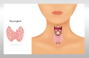
The gland is located in the middle of the lower neck, below the larynx (voice box) and wraps around the front half of the trachea (windpipe). It is shaped like a bow tie, just above the collarbones, having two halves (lobes) joined by a small tissue bar (isthmus.). You can’t always feel a normal gland.
The most common diseases of the thyroid gland are an overactive gland (hyperthyroidism), an under-active gland (hypothyroidism ), and thyroid enlargement due to over-activity (as in Graves’ disease), from under-activity (as in hypothyroidism), or from tumors. An enlarged thyroid gland is often called a “goiter.”
An abnormal thyroid is usually detected by a lump or a mass on the neck, which can be benign or malignant and may appear gradually or rapidly. However, many patients may present with a single enlarged thyroid mass. Other indicators are difficulty breathing or swallowing.
The diagnosis of a thyroid function abnormality or a thyroid mass is made by taking a medical history and a physical examination (Hearing testing). In addition, blood tests and imaging studies or fine-needle aspiration may be required. As part of the exam, your doctor will examine your neck and ask you to lift up your chin to make your thyroid gland more prominent. You may be asked to swallow during the examination, which helps to feel the thyroid and any mass in it. Tests your doctor may order include:
Evaluation of the larynx/vocal cords with a mirror or a fiberoptic telescope
An ultrasound examination of your neck and thyroid
Blood tests of thyroid function
A radioactive thyroid scan
A fine-needle aspiration biopsy
A chest X-ray
A CT or MRI scan
ENT of NJ offers a full range of same day minimally invasive and diagnostics treatments, including thyroid ultrasounds, ultrasound guided fine needle biopsy, and surgical removal utilizing small incisions to improve recovery times.
* Also, a nerve monitor is used in the procedure to gauge and protect the voice nerve during surgery.
*Some or all of the information found here can be attributed to The American Academy of Otolaryngology website